Vagus Nerve Stimulation for Pain: What the Research Shows
Introduction: Pain as a Neuromodulation Target
Chronic pain affects an estimated 1.5 billion people worldwide and remains one of the most significant unmet needs in modern medicine. Despite decades of pharmacological development, current approaches to pain management carry substantial limitations. Opioid analgesics, while effective for acute pain, are associated with tolerance, dependence, and a well-documented epidemic of misuse. Non-steroidal anti-inflammatory drugs (NSAIDs) carry cardiovascular and gastrointestinal risks with long-term use. Anticonvulsants and antidepressants repurposed for neuropathic pain offer modest efficacy and often produce intolerable side effects.
These limitations have driven growing interest in neuromodulation — the use of electrical or magnetic stimulation to alter neural activity — as an alternative or complementary strategy for pain management. Among the neuromodulation approaches under investigation, vagus nerve stimulation (VNS) has emerged as one of the most actively researched, supported by a convergence of preclinical evidence, mechanistic plausibility, and early clinical data.
The rationale for targeting the vagus nerve in pain is grounded in foundational neuroscience. The vagus nerve — the longest cranial nerve in the body — carries both afferent (sensory) and efferent (motor) fibres that connect peripheral organs to key brainstem nuclei involved in pain processing. Electrical stimulation of vagal afferents has been shown in animal models to activate the descending pain modulatory system, a network of brainstem and midbrain structures that can suppress nociceptive (pain-signalling) transmission at the level of the spinal cord and trigeminal nucleus.
This article reviews the neuroanatomical basis for vagal pain modulation, the clinical evidence across specific pain conditions — particularly migraine and fibromyalgia — and the current limitations of this rapidly evolving field.
Neuroanatomical Basis: How the Vagus Nerve Connects to Pain Processing
Understanding why VNS may influence pain requires tracing the anatomical pathway from vagal afferent fibres to the brain regions that govern pain perception and modulation.
The Nucleus Tractus Solitarius: The First Relay
Approximately 80% of vagal fibres are afferent, carrying sensory information from the viscera to the brain. These fibres terminate primarily in the nucleus tractus solitarius (NTS) in the dorsomedial brainstem. The NTS serves as the critical first relay station for vagal input and has extensive projections to higher brain structures involved in pain processing (Randich & Gebhart, 1992).
From the NTS, vagal afferent signals are distributed to several key nodes in the pain network.
The Locus Coeruleus and Noradrenergic Modulation
The NTS projects directly to the locus coeruleus (LC), the brain's principal noradrenergic nucleus. The LC plays a central role in arousal, attention, and — critically — descending pain inhibition. Noradrenaline released from LC projections to the spinal dorsal horn activates alpha-2 adrenergic receptors on second-order nociceptive neurons, directly inhibiting pain transmission (Randich & Gebhart, 1992).
This NTS-LC-noradrenaline pathway is considered one of the primary mechanisms through which VNS may produce analgesic effects. Animal studies have demonstrated that VNS enhances tonic noradrenergic output from the LC while modulating phasic responses, potentially establishing a more stable analgesic baseline (Manta et al., 2009).
The Periaqueductal Grey and Descending Inhibition
The periaqueductal grey (PAG) in the midbrain is the master control centre of the endogenous descending pain modulatory system. The PAG integrates input from cortical, limbic, and brainstem sources and projects to the rostral ventromedial medulla (RVM), which in turn sends descending projections to the spinal dorsal horn and trigeminal nucleus caudalis. These descending pathways can either facilitate or inhibit nociceptive transmission, depending on the balance of "on-cell" and "off-cell" activity in the RVM.
Functional neuroimaging studies have demonstrated that transcutaneous auricular VNS (taVNS) modulates functional connectivity of the PAG in migraine patients. Zhang et al. (2021) showed that 1 Hz taVNS specifically enhanced connectivity between the PAG and the medial cingulate cortex, anterior cingulate cortex, and anterior insula — all regions involved in the affective and evaluative dimensions of pain. This frequency-dependent modulation of PAG connectivity provides direct neuroimaging evidence that taVNS engages the descending pain modulatory system in humans.
Serotonergic and GABAergic Contributions
The NTS also projects to the raphe nuclei — the brainstem's serotonergic centres — which contribute to descending pain inhibition through serotonin release in the spinal cord. Additionally, VNS has been shown to increase levels of gamma-aminobutyric acid (GABA) — the brain's primary inhibitory neurotransmitter — in the central nervous system (Ben-Menachem et al., 1995). Since GABAergic deficits have been implicated in central sensitisation (the pathological amplification of pain signalling), VNS-mediated increases in GABA may help restore inhibitory tone in pain circuits.
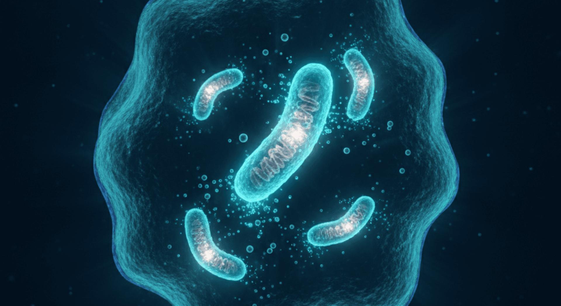
The Cholinergic Anti-Inflammatory Pathway
Beyond direct neural modulation, VNS activates the cholinergic anti-inflammatory pathway (CAP) — a vagal efferent circuit that suppresses the production of pro-inflammatory cytokines including TNF-alpha, IL-1beta, and IL-6 (Tracey, 2002). Since neuroinflammation and peripheral inflammation are increasingly recognised as contributors to chronic pain states, the anti-inflammatory properties of VNS may represent an additional analgesic mechanism, particularly in conditions such as fibromyalgia and inflammatory arthritis where persistent inflammation drives pain sensitisation.
Migraine and Headache: The Most Developed Clinical Evidence
Migraine represents the pain condition for which VNS has the most extensive clinical evidence, and it is the first pain indication for which a non-invasive VNS device has received regulatory clearance.
Early Observations and Preclinical Foundation
The link between VNS and headache relief was first observed incidentally in epilepsy patients with implanted VNS devices who reported reductions in migraine frequency and severity. These clinical observations were supported by preclinical work demonstrating that VNS could suppress trigeminal nociceptive processing — the neural pathway central to migraine pathophysiology.
Bohotin et al. (2003) provided key preclinical evidence, demonstrating that left cervical VNS in awake rats reduced formalin-induced nociceptive behaviour by 60–96% and significantly attenuated Fos-immunoreactivity in the trigeminal nucleus caudalis (TNC) — a marker of nociceptive neuronal activation. When the direct VNS effect on Fos expression was subtracted, the reduction of formalin-induced nociceptor activation was approximately 55%.
Building on this, Oshinsky et al. (2014) demonstrated that non-invasive VNS reversed trigeminal allodynia in rats exposed to repeated inflammatory dural stimulation and identified the mechanism: nVNS reduced the pathological elevation of extracellular glutamate in the TNC by approximately 70%, bringing levels back to those observed in naive control animals. These findings pointed to glutamate modulation as a key mechanism through which VNS may treat migraine-related trigeminal pain.
Acute Migraine Treatment: The PRESTO Trial
The most rigorous evidence for non-invasive VNS (nVNS) in acute migraine comes from the PRESTO trial (Tassorelli et al., 2018) — a multicentre, randomised, double-blind, sham-controlled study of 248 patients with episodic migraine.
Participants self-administered cervical nVNS (gammaCore) or sham stimulation within 20 minutes of migraine onset. The results demonstrated that nVNS was superior to sham for pain freedom at 30 minutes (12.7% vs 4.2%; p = 0.012) and 60 minutes (21.0% vs 10.0%; p = 0.023). At 120 minutes — the primary endpoint — the difference favoured nVNS but did not reach statistical significance (30.4% vs 19.7%; p = 0.067). A post hoc repeated-measures analysis across all time points confirmed a significant overall treatment effect (OR 2.3; 95% CI 1.2–4.4; p = 0.012).
The PRESTO trial provided Class I evidence supporting nVNS for the acute treatment of episodic migraine, though the effect sizes were modest compared to oral triptans.
The Pilot Study: Goadsby et al. (2014)
Prior to PRESTO, an open-label pilot study by Goadsby et al. (2014) assessed transcutaneous cervical VNS in 19 patients with moderate-to-severe migraine. Two-hour pain-free response was 21%, and pain relief (reduction to mild or no pain) was achieved in 47% of patients. While limited by the lack of a sham control, these results provided early proof-of-concept for nVNS as an acute migraine treatment and informed the design of subsequent controlled trials.
Migraine Prevention: The EVENT Study
Silberstein et al. (2016) conducted the EVENT study — the first multicentre, double-blind, sham-controlled pilot study of nVNS for chronic migraine prevention. Patients with chronic migraine (15 or more headache days per month) self-administered cervical nVNS or sham treatment three times daily for two months. The primary endpoints were safety and tolerability, which were confirmed. While the study was not powered for definitive efficacy conclusions, the results provided the foundation for the larger PREMIUM II prevention trial.
Auricular taVNS for Chronic Migraine
Straube et al. (2015) conducted a randomised, monocentric trial comparing 1 Hz versus 25 Hz auricular taVNS for chronic migraine prevention. In the per-protocol analysis, patients receiving 1 Hz stimulation showed a significantly greater reduction in headache days per 28 days compared to the 25 Hz group. This finding aligns with the neuroimaging evidence from Zhang et al. (2021) showing that 1 Hz taVNS more effectively modulates PAG connectivity than higher-frequency stimulation, suggesting that lower frequencies may be preferentially analgesic.
FDA Clearance
In January 2018, the gammaCore device (electroCore, LLC) received FDA 510(k) clearance for the acute treatment of pain associated with migraine in adult patients — making it the first non-invasive VNS device cleared for a pain indication. The device had previously received clearance for episodic cluster headache in 2017, based on the ACT1 trial (Silberstein et al., 2016b), which demonstrated that nVNS achieved pain relief in 34.2% of episodic cluster headache patients compared to 10.6% with sham (p = 0.008). gammaCore has since received six FDA-cleared indications for headache disorders, including both acute and preventive treatment of migraine.
Fibromyalgia: Emerging Evidence
Fibromyalgia syndrome (FMS) is a chronic widespread pain condition characterised by central sensitisation, autonomic dysfunction, and dysregulated pain processing. The involvement of both autonomic imbalance and central pain amplification makes fibromyalgia a theoretically compelling target for VNS.
Kutlu et al. (2020): The First Randomised Trial
The most significant study to date on VNS for fibromyalgia was conducted by Kutlu et al. (2020). In this pilot randomised controlled trial, 60 female patients diagnosed with FMS (according to ACR 2010 criteria) were randomly divided into two groups: one receiving 20 sessions of auricular VNS combined with a home-based exercise programme over four weeks, and the other receiving the exercise programme alone.
Both groups improved significantly in pain (Visual Analogue Scale), anxiety (Beck Anxiety Scale), depression (Beck Depression Scale), and quality of life. However, the taVNS group showed additional improvements in select domains of the Short Form-36 (SF-36), including physical function, social functioning, and pain. Although between-group differences did not reach statistical significance overall, the taVNS group showed consistently larger gains across outcomes.
The study had important limitations: it was relatively small (n = 60), did not include a sham stimulation control (comparing taVNS plus exercise to exercise alone), and had a short follow-up period. Nonetheless, it represented the first randomised investigation of VNS for fibromyalgia and provided pilot data supporting larger, sham-controlled trials. For a dedicated, up-to-date review of this evidence, see VNS for fibromyalgia.
Mechanistic Rationale for VNS in Fibromyalgia
The theoretical case for VNS in fibromyalgia rests on several converging mechanisms:
- Central sensitisation — Fibromyalgia is characterised by augmented central pain processing and impaired descending pain inhibition. By activating the PAG-RVM descending inhibitory system, VNS may help restore the inhibitory tone that is deficient in FMS
- Autonomic dysfunction — Patients with fibromyalgia consistently show reduced heart rate variability (HRV) and sympathovagal imbalance. VNS directly addresses this by increasing parasympathetic activity
- Neuroinflammation — Emerging evidence suggests that neuroinflammation, including elevated pro-inflammatory cytokines and activated glial cells, contributes to central sensitisation in FMS. The anti-inflammatory properties of VNS via the cholinergic anti-inflammatory pathway may be relevant here
- Sleep disruption — Non-restorative sleep is a hallmark of FMS. VNS has been shown to improve sleep architecture in other populations, potentially addressing another contributor to pain amplification
Chronic Pain Mechanisms: Beyond Specific Conditions
Experimental Pain Studies in Healthy Volunteers
Before examining clinical pain populations, researchers established that VNS can modulate pain perception in controlled experimental settings.
Kirchner et al. (2000) conducted one of the earliest human studies, examining pain thresholds in 10 epilepsy patients before and after implantation of a VNS device. After 8–14 weeks of stimulation, patients showed decreased temporal summation of pain (wind-up) and reduced tonic pressure pain — critically, this effect was independent of the VNS on/off cycle, suggesting a chronic reorganisation of central pain processing rather than an acute, stimulus-locked effect. This pattern of gradual, lasting change is consistent with the broader evidence that VNS promotes neuroplastic remodelling of neural circuits over time.
Busch et al. (2013) extended these findings to non-invasive stimulation, demonstrating in a randomised, sham-controlled crossover study of 48 healthy volunteers that transcutaneous auricular VNS increased mechanical and pressure pain thresholds and reduced pain ratings during a sustained tonic heat paradigm. Importantly, VNS selectively modulated pain-specific processing without altering detection thresholds for innocuous touch, warmth, or cold — suggesting a targeted analgesic mechanism rather than generalised sensory blunting.
Napadow et al. (2012) introduced a novel approach — respiratory-gated auricular vagal afferent nerve stimulation (RAVANS) — and tested it in patients with chronic pelvic pain. A single session of RAVANS significantly reduced evoked pain intensity and temporal summation of mechanical pain compared to non-vagal auricular stimulation, along with a significant anxiolytic effect. The respiratory gating was designed to optimise vagal afferent activation by synchronising stimulation with exhalation, when vagal tone is naturally highest.
The Anti-Inflammatory Dimension of Pain
Chronic pain is increasingly understood not solely as a neural phenomenon but as a neuroimmune process. Persistent inflammation — whether peripheral, central, or both — drives and maintains pain sensitisation through multiple mechanisms: sensitisation of peripheral nociceptors, activation of spinal microglia and astrocytes, and disruption of the blood-brain barrier.
The vagus nerve's capacity to suppress inflammation via the cholinergic anti-inflammatory pathway (CAP) therefore represents a distinct analgesic mechanism. By reducing circulating levels of TNF-alpha, IL-1beta, and IL-6, VNS may address the inflammatory component of chronic pain that is not targeted by most conventional analgesics. This mechanism may be particularly relevant in conditions where inflammation and pain coexist, including inflammatory arthritis, inflammatory bowel disease, and post-surgical pain.
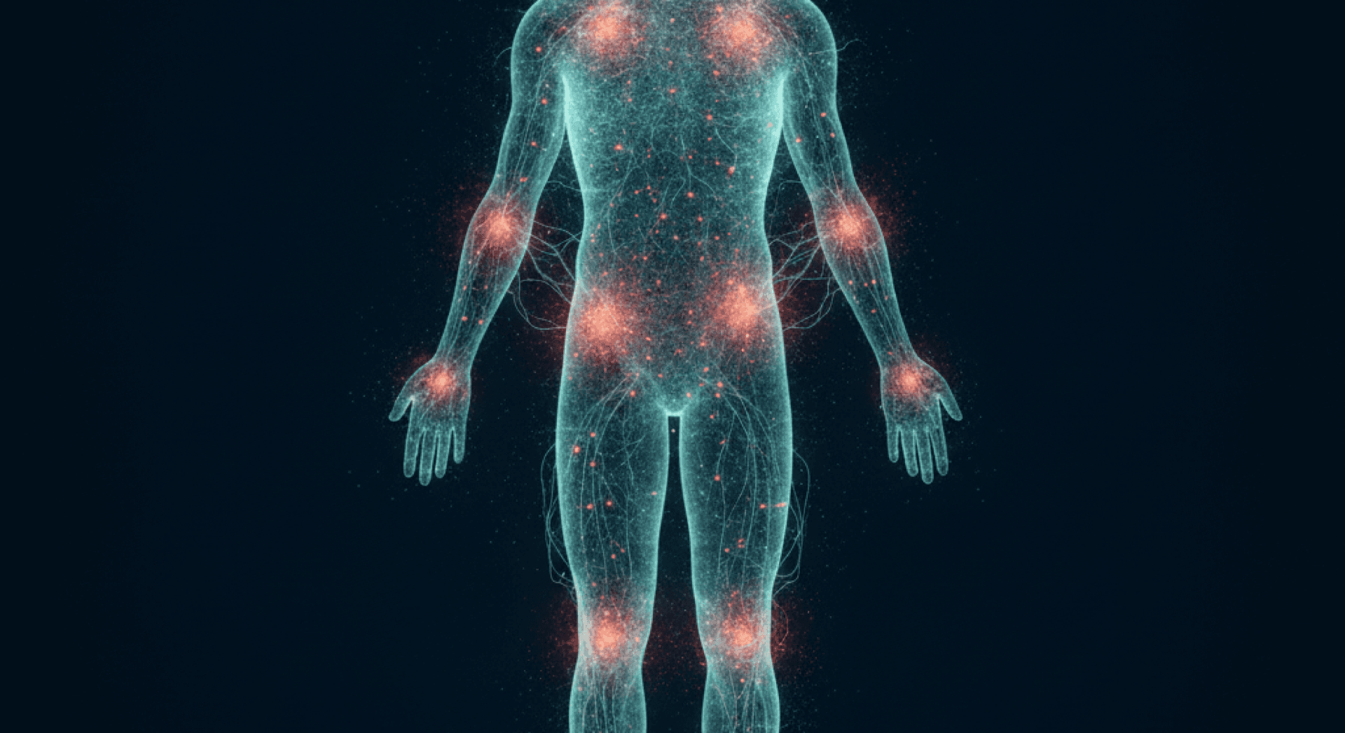
Autonomic Rebalancing and the Pain-Stress-Autonomic Axis
Chronic pain is almost invariably accompanied by autonomic dysfunction — typically manifested as sympathetic overactivation and parasympathetic withdrawal. This autonomic imbalance is not merely a consequence of pain; it actively perpetuates pain through several mechanisms:
- Sympathetic activation increases muscle tension and vasoconstriction, contributing to ischaemic pain
- Reduced parasympathetic activity impairs the body's capacity for recovery and tissue repair
- Autonomic dysregulation disrupts sleep, amplifying pain sensitisation through sleep deprivation
- The resulting stress response elevates cortisol and inflammatory mediators, further fuelling the pain cycle
By increasing vagal tone and shifting the autonomic balance toward parasympathetic dominance, VNS may help interrupt this self-reinforcing cycle. This autonomic rebalancing effect — consistently demonstrated through improvements in heart rate variability — may represent one of the most broadly applicable mechanisms of VNS for chronic pain, regardless of the specific underlying condition.
Clinical Limitations and Methodological Challenges
While the evidence base for VNS in pain management is growing, several significant limitations must be acknowledged.
Small Sample Sizes
With the exception of the PRESTO trial (n = 248) and ACT1 (n = 150), most studies of VNS for pain have employed small samples — often fewer than 50 participants. The Kutlu et al. (2020) fibromyalgia trial included 60 participants, and the Kirchner et al. (2000) and Napadow et al. (2012) studies had 10 and 15 participants respectively. Small samples increase the risk of both false-positive and false-negative findings and limit the generalisability of results.
Parameter Variability
There is no consensus on optimal stimulation parameters for pain. Studies vary widely in:
- Frequency: 1 Hz, 20 Hz, 25 Hz, and other frequencies have been used, with emerging evidence suggesting that lower frequencies (1 Hz) may be more effective for engaging descending pain inhibition
- Intensity: Ranges from sub-perceptual to the maximum tolerable level
- Duration: Single 2-minute stimulations to 4-hour daily sessions
- Stimulation site: Cervical (gammaCore), auricular cymba conchae (taVNS), and auricular tragus stimulation all target vagal afferents but may activate different fibre populations with different efficacy
- Duty cycle and session frequency: Daily, multiple times daily, or intermittent protocols have all been employed
This heterogeneity makes it difficult to compare findings across studies and to identify which protocols are most effective for which pain conditions.
Sham Stimulation Challenges
Designing a credible and truly inert sham condition for VNS trials remains methodologically challenging. Most taVNS studies use earlobe stimulation as a sham, based on the understanding that the earlobe is innervated by the great auricular nerve rather than the vagus nerve. However, debate persists about whether earlobe stimulation is completely inert — some evidence suggests it may produce mild physiological effects, potentially reducing the apparent superiority of active stimulation (Badran et al., 2018).
For cervical nVNS, sham devices that are identical in appearance and produce similar sensory feedback (tingling) but do not deliver therapeutic electrical stimulation have been developed, though ensuring effective blinding remains a concern.
Short Follow-Up Periods
Most clinical trials have assessed outcomes over days to weeks. The durability of VNS-induced analgesia — whether pain relief persists after stimulation is discontinued, and whether long-term daily use produces sustained benefits or leads to tolerance — remains largely unknown. The chronic reorganisation of pain processing observed by Kirchner et al. (2000) is suggestive of lasting effects, but this needs confirmation in larger studies with longer follow-up.
Lack of Head-to-Head Comparisons
Few studies have directly compared VNS to established pain treatments. The PRESTO trial results, while statistically significant, showed pain-free rates that are lower than those typically reported for triptans in acute migraine. Without direct comparisons, it is difficult to position VNS within the existing treatment hierarchy for any pain condition.
Condition-Specific Evidence Gaps
Beyond migraine and cluster headache, the evidence for VNS in specific pain conditions remains at the pilot or proof-of-concept stage. For fibromyalgia, neuropathic pain, low back pain, and visceral pain, the data are too limited to draw firm conclusions about efficacy. Large, condition-specific trials with appropriate sham controls are needed.
Conclusion
The evidence linking vagus nerve stimulation to pain modulation rests on a solid neuroanatomical and mechanistic foundation. The vagus nerve, through its projections from the NTS to the locus coeruleus, periaqueductal grey, and raphe nuclei, is anatomically positioned to engage the full descending pain modulatory system. Preclinical studies have consistently demonstrated that VNS can suppress nociceptive processing, and experimental pain studies in humans have confirmed that both invasive and transcutaneous VNS can increase pain thresholds and reduce pain perception.
For migraine, the clinical evidence has progressed to the stage of randomised, sham-controlled trials and regulatory clearance, making it the most validated pain indication for non-invasive VNS. For fibromyalgia and other chronic pain conditions, the evidence remains at an earlier stage — encouraging, mechanistically plausible, but requiring confirmation in larger and more rigorous trials.
What makes VNS particularly compelling as a pain management approach is the convergence of multiple analgesic mechanisms: direct activation of descending pain inhibition, anti-inflammatory effects via the cholinergic anti-inflammatory pathway, autonomic rebalancing, and modulation of central sensitisation. Few other interventions simultaneously address this many dimensions of the chronic pain experience.
The field is still maturing. Optimal stimulation parameters, condition-specific protocols, predictors of individual response, and long-term outcomes all require further investigation. However, the trajectory of evidence — from foundational neuroscience through preclinical studies to clinical trials — provides a coherent and increasingly substantive case for VNS as a meaningful addition to the pain management toolkit.
---
References
Badran, B.W. et al. (2018). Neurophysiologic effects of transcutaneous auricular vagus nerve stimulation (taVNS) via electrical stimulation of the tragus. Brain Stimulation, 11(3), 492–500.
Ben-Menachem, E. et al. (1995). Effects of vagus nerve stimulation on amino acids and other metabolites in the CSF of patients with partial seizures. Epilepsy Research, 20(3), 221–227.
Bohotin, C. et al. (2003). Vagus nerve stimulation in awake rats reduces formalin-induced nociceptive behaviour and fos-immunoreactivity in trigeminal nucleus caudalis. Pain, 101(1–2), 3–12.
Busch, V. et al. (2013). The effect of transcutaneous vagus nerve stimulation on pain perception — an experimental study. Brain Stimulation, 6(2), 202–209.
Goadsby, P.J. et al. (2014). Effect of noninvasive vagus nerve stimulation on acute migraine: an open-label pilot study. Cephalalgia, 34(12), 986–993.
Kirchner, A. et al. (2000). Left vagus nerve stimulation suppresses experimentally induced pain. Neurology, 55(8), 1167–1171.
Kutlu, N. et al. (2020). The impact of auricular vagus nerve stimulation on pain and life quality in patients with fibromyalgia syndrome. BioMed Research International, 2020, 8656218.
Manta, S. et al. (2009). Enhancement of the function of rat serotonin and norepinephrine neurons by sustained vagus nerve stimulation. Journal of Psychiatry & Neuroscience, 34(4), 272–280.
Napadow, V. et al. (2012). Evoked pain analgesia in chronic pelvic pain patients using respiratory-gated auricular vagal afferent nerve stimulation. Pain Medicine, 13(6), 777–789.
Oshinsky, M.L. et al. (2014). Noninvasive vagus nerve stimulation as treatment for trigeminal allodynia. Pain, 155(5), 1037–1042.
Randich, A. & Gebhart, G.F. (1992). Vagal afferent modulation of nociception. Brain Research Reviews, 17(2), 77–99.
Silberstein, S.D. et al. (2016a). Chronic migraine headache prevention with noninvasive vagus nerve stimulation: the EVENT study. Neurology, 87(5), 529–538.
Silberstein, S.D. et al. (2016b). Non-invasive vagus nerve stimulation for the acute treatment of cluster headache: findings from the randomized, double-blind, sham-controlled ACT1 study. Headache, 56(8), 1317–1332.
Straube, A. et al. (2015). Treatment of chronic migraine with transcutaneous stimulation of the auricular branch of the vagal nerve (auricular t-VNS): a randomized, monocentric clinical trial. The Journal of Headache and Pain, 16, 543.
Tassorelli, C. et al. (2018). Noninvasive vagus nerve stimulation as acute therapy for migraine: the randomized PRESTO study. Neurology, 91(4), e364–e373.
Tracey, K.J. (2002). The inflammatory reflex. Nature, 420(6917), 853–859.
Zhang, Y. et al. (2021). Different modulation effects of 1 Hz and 20 Hz transcutaneous auricular vagus nerve stimulation on the functional connectivity of the periaqueductal gray in patients with migraine. Journal of Translational Medicine, 19, 354.